
Light and Specular Microscopy of the Cornea: A Comprehensive Exploration

The cornea, a transparent and curved outermost layer of the eye, plays a crucial role in focusing light onto the retina for clear vision. Understanding its intricate structure and composition is essential for diagnosing and treating corneal diseases. Light and specular microscopy are two essential techniques employed to examine the cornea at different depths and scales. This article delves into the principles, applications, and clinical significance of these microscopy techniques, providing a comprehensive overview for eye care professionals.
Light Microscopy of the Cornea
Light microscopy utilizes visible light to visualize the cornea's cellular and histological features. There are two main types of light microscopy commonly used in corneal examination:

5 out of 5
| Language | : | English |
| File size | : | 9514 KB |
| Text-to-Speech | : | Enabled |
| Screen Reader | : | Supported |
| Enhanced typesetting | : | Enabled |
| Print length | : | 238 pages |
a) Histological Light Microscopy
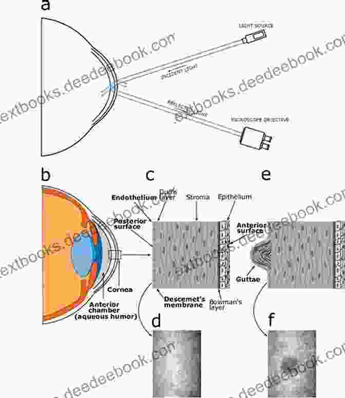
In histological light microscopy, a thin section of the cornea is stained and mounted on a glass slide. This technique provides detailed information about the cornea's overall architecture, cell types, and pathological changes. It enables the identification of corneal layers, such as the epithelium, Bowman's layer, stroma, Descemet's membrane, and endothelium. Histological light microscopy is particularly useful in diagnosing corneal infections, scarring, and degenerative conditions.

b) Confocal Light Microscopy
Confocal light microscopy employs a laser scanning device to generate high-resolution images of the cornea. It provides optical sections at different depths, allowing for detailed visualization of corneal structures in three dimensions. Confocal light microscopy is particularly useful for assessing corneal thickness, epithelial integrity, and nerve fiber density. It has applications in diagnosing corneal dystrophies, inflammatory diseases, and refractive surgical complications.

Specular Microscopy of the Cornea
Specular microscopy is a non-invasive technique that utilizes reflected light to examine the corneal endothelium, the innermost cell layer of the cornea. It provides valuable information about endothelial cell density, size, and morphology.
Specular microscopy is primarily used for:
a) Endothelial Cell Count
Endothelial cell count provides an indirect measure of corneal health. A healthy cornea typically has a high density of polygonal-shaped endothelial cells. Specular microscopy allows the quantification of endothelial cell density, which can decrease with age, trauma, and surgical interventions.
b) Endothelial Cell Morphology
Specular microscopy enables the visualization of endothelial cell shape, size, and regularity. Abnormal endothelial cell morphology, such as polymegathism (enlarged cells) or pleomorphism (cells with irregular shapes),can indicate underlying corneal diseases or surgical complications.
c) Corneal Edema Detection
Excessive fluid accumulation in the cornea, known as corneal edema, can be detected using specular microscopy. Edema appears as a hazy or cloudy appearance on specular images. It can be caused by various factors, including endothelial damage, inflammation, and glaucoma.
Applications in Clinical Practice
Light and specular microscopy are indispensable tools for diagnosing and managing a wide range of corneal conditions, including:
a) Infectious Keratitis
Histological light microscopy is essential for diagnosing infectious keratitis by identifying the causative microorganisms. Confocal light microscopy can provide additional information about the depth of corneal involvement and guide treatment decisions.
b) Corneal Dystrophies
Light and specular microscopy are used to characterize different types of corneal dystrophies, which are inherited disorders that affect the cornea's structure and function.
c) Corneal Scars
Histological light microscopy helps assess the extent and nature of corneal scarring, while confocal light microscopy can determine the depth and cellular composition of the scar tissue.
d) Postoperative Evaluation
Specular microscopy is critical for monitoring corneal endothelial cell health after refractive surgeries, such as LASIK and PRK. It can detect endothelial cell loss and guide decisions regarding future surgical interventions.
e) Contact Lens Evaluation
Specular microscopy can evaluate the impact of contact lens wear on corneal endothelium. It can detect changes in endothelial cell morphology and density, which may influence contact lens tolerance and long-term corneal health.
Future Directions
Advances in microscopy technology are constantly improving the capabilities of corneal imaging. Recent developments include:
a) In Vivo Confocal Microscopy
In vivo confocal microscopy enables real-time imaging of the cornea, providing dynamic information about cellular and structural changes. It has applications in diagnosing early corneal diseases and monitoring treatment response.
b) Adaptive Optics Microscopy
Adaptive optics microscopy corrects corneal aberrations, allowing for high-resolution imaging of deeper corneal layers. It has potential applications in studying the ultrastructure of the corneal stroma and diagnosing subtle corneal abnormalities.
Light and specular microscopy techniques provide essential insights into the structure and health of the cornea. Histological light microscopy allows for detailed histological examination, while confocal light microscopy offers high-resolution imaging of corneal layers in three dimensions. Specular microscopy is specialized for examining the corneal endothelium, providing valuable information about cell density, morphology, and corneal edema. These microscopy techniques are crucial for diagnosing and managing a wide range of corneal diseases, guiding treatment decisions, and advancing our understanding of corneal structure and function. With continued advancements in microscopy technology, we can expect even more refined and informative corneal imaging in the future.
5 out of 5
| Language | : | English |
| File size | : | 9514 KB |
| Text-to-Speech | : | Enabled |
| Screen Reader | : | Supported |
| Enhanced typesetting | : | Enabled |
| Print length | : | 238 pages |
Do you want to contribute by writing guest posts on this blog?
Please contact us and send us a resume of previous articles that you have written.
 Book
Book Novel
Novel Page
Page Chapter
 Text
Text Genre
Genre Paperback
 E-book
 Magazine
 Sentence
 Bibliography
 Preface
 Synopsis
 Bestseller
 Classics
 Library card
 Biography
 Reference
 Encyclopedia
 Narrator
 Character
 Librarian
 Catalog
 Card Catalog
 Borrowing
 Stacks
 Archives
 Academic
 Reading Room
 Rare Books
 Special Collections
 Interlibrary
 Literacy
 Study Group
 Dissertation
 Storytelling
 Awards
 Reading List
 Book Club
 Theory
 Denise Mina
 Wilfried Achenbach
 Jeannie Pitt
 John Maynard Keynes
 Moazzam Begg
 Te Wu
Te Wu Sherry Rossman
 Joseph Lelyveld
 Sandra Staines
 Michael Avon Oeming
 Melissa Nobles
 Jan Richardson
 Macartan Humphreys
 Michael Hodgson
 Elizabeth St John
 R L Margolin
 A Tebbs
 1st Ed 2017 Edition Kindle Edition
 Paul Ireland
 Tom Pearson
Light bulbAdvertise smarter! Our strategic ad space ensures maximum exposure. Reserve your spot today!


 Bryce FosterMemoirs of Filipino American Scott and Laurie Oki: A Rich Tapestry of Asian...
 Harold PowellUnraveling the Pullman Hilton Christmas Mystery: A Journey Through Time and...
 Fyodor DostoevskyFollow ·4.8k
 Oscar WildeFollow ·2.8k
 Dan HendersonFollow ·13.4k
 Colin RichardsonFollow ·12.6k
 Aron CoxFollow ·19.5k
 Jacob FosterFollow ·19.2k
 Christopher WoodsFollow ·6.4k
 Benji PowellFollow ·6.3k

 Elton Hayes
Unveiling the Enchanting Legends of Emelina Grace and...
Emelina Grace: The...

 Evan Simmons
What If Vietnam Never Happened: Foresight and Hindsight...
Published in 1955, Graham Greene's The Quiet...

 Dave Simmons
 Camden Mitchell
The Rise of Specialty Coffee, Craft Beer, Vegan Food,...
In recent years,...

 Corey Hayes
Modern Project Creative Techniques: A Comprehensive Guide...
In today's competitive business landscape,...

 Norman Butler
5 out of 5
| Language | : | English |
| File size | : | 9514 KB |
| Text-to-Speech | : | Enabled |
| Screen Reader | : | Supported |
| Enhanced typesetting | : | Enabled |
| Print length | : | 238 pages |
